
According to the International Association of Dental Traumatology (IADT), a third of all adults will experience some type of trauma to their permanent teeth in their lifetime, the most common of which will be to the crown of a tooth.
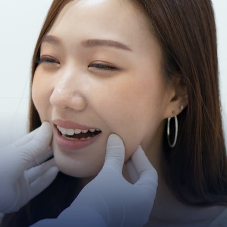
Knowing what to do when visited by a patient who has experienced a traumatic dental injury (TDI) is key to increase the likelihood your patient will experience a favourable treatment outcome. To accomplish this, correct diagnosis, treatment, and follow up are critical.

CVOS Oral Surgery Partners With Dentists
A goal of CVOS Oral Surgery is to fully support our referring dentists in whatever ways we can for the benefit of the patient…
…And, in most cases, patients who have suffered a TDI will seek primary care from their dentist.
Therefore, to aid in the diagnosis and treatment of TDIs at the time of, or immediately following an injury, below is a summary of the IADT guidelines (go here to access the full guidelines).
Summary Guidelines for Fractures of Teeth and Alveolar Bone
The information presented below is a summary of the IADT Guidelines and meant for reference only.
Infraction
An infraction results when the enamel of the tooth is cracked, but the structure of the tooth is retained and there is generally little to no tenderness.
In these cases, a periapical x-ray is recommended, and no abnormalities should be visible.
Treatment can range from no treatment to etching and sealing to prevent discolouration and little or no follow up is required.
Enamel Fracture
A complete fracture resulting in the loss of a tooth’s enamel, but no visible loss of dentin indicates an enamel fracture.
In this case, the IADT recommends periapical, occlusal and eccentric radiographs be taken. While it is expected that the loss of enamel will be evident, these additional scans (vs. a standard infraction) are recommended to rule out a more severe diagnosis. Additional imaging may also be required to locate tooth fragments in the lip or cheek.
Treatment is straightforward. If the tooth fragment is available, it can be bonded to the tooth in question. However, depending on the severity and location of the fracture, additional restorative work may be called for.
Enamel-Dentin Fracture
This TDI can be clinically diagnosed when the fracture is limited to the enamel and dentin and there is a loss of tooth structure, but the pulp is not exposed. A percussion and sensibility test can be used to supplement the diagnosis.
It is also recommended that x-rays be taken using the periapical and occlusal views to rule out a root fracture or tooth displacement. Images of the cheek or lip may also be beneficial to locate tooth fragments.
In these cases, it is always ideal to bond the tooth fragment when possible. However, if this isn’t possible, a provisional treatment such as covering the exposed dentin with glass ionomer (after covering the dentin with a calcium hydroxide base if the exposed dentin is within 0.5mm of the pulp) or a more permanent bonding agent and composite resin should be performed.
Enamel-Dentin-Pulp Fracture
This TDI is diagnosed when there is evidence that the fracture in the enamel and dentin has exposed the pulp. Like the enamel-dentin fracture, a percussion test can be completed to evaluate tenderness and exposed pulp is likely to be sensitive to stimuli.
Scanning the area affected by the injury using the 3 previously mentioned views, the enamel-dentin loss will be visible.
For treatment in younger patients, it is preferable to preserve the pulp vitality by pulp capping or a carrying out a partial pulpotomy, using calcium hydroxide over the wound. In more mature patients, a root canal is typically the recommended treatment. Of course, as with the previous TDIs, where the tooth fragment is available, bonding it back on to the tooth is ideal.
With this particular type of TDI, further future restoration may be an option.
Crown-Root Fracture Without Pulp Exposure
A more severe injury than those previously discussed, upon clinical examination, this injury involves a fracture in the enamel, dentin, and cementum with the loss of tooth structure but no exposed pulp. In this case, the fracture in the tooth extends below the gum line (gingival margin). A percussion test will reveal tenderness and a sensibility test will be positive for an apical fragment. There may also be mobility in the coronal fragment.
Periapical, occlusal, and eccentric x-rays are suggested to detect fracture lines in the root.
With this type of injury, emergency treatment such as the temporary stabilization of the loose portion to the adjacent teeth may be warranted.
When treatment is not an emergency, several alternatives exist – from removal of the fragment only to the removal of the coronal crown-root segment. In the latter case, the removal of the fragment may be followed by subsequent restoration work, endodontic treatment, orthodontic extrusion of the root, and/or the surgical repositioning of the root. A gingivectomy, ostectomy, and/or osteoplasty may also be recommended.
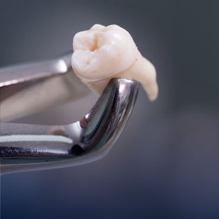
Once the fragment has been removed, the correct implant solution can be planned for the patient to follow the extraction.
Crown-Root Fracture with Pulp Exposure
This most severe of listed fractures reveals itself upon examination as a fracture of the enamel, dentin, and cementum with exposed pulp. A percussion test will reveal tenderness and there will be mobility in the coronal fragment.
The clinical findings can be supplemented by carrying out x-rays of the periapical and occlusal views.

Emergency treatment includes the stabilization of the loose fragment to adjacent teeth. It is also desirable to preserve pulp vitality via a partial pulpotomy. In mature patients, root canal treatment may also be the preferred course of action.
The non-emergency treatment options vary depending on the severity of the injury. It is strongly recommended that a practitioner refer to the IADT guidelines and consult with a surgeon where appropriate to increase the likelihood of a favourable patient outcome.
Optimizing Patient Care
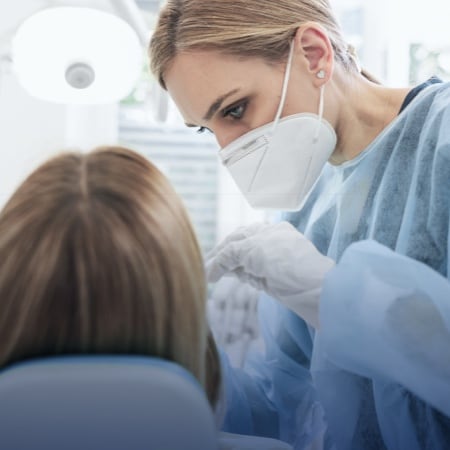
Most people, when suffering from a TDI that doesn’t require hospitalization will first seek treatment from their family dentist, who is in the best position to provide immediate and/or urgent care to the patient.
However, for more severe injuries, it is likely that additional treatment may be required by specialists such as CVOS Oral Surgery’s team of oral & maxillofacial surgeons.
CVOS Oral Surgery has a long history of partnering with dentists to ensure their patients receive the scope of care needed to address the trauma and ultimately recover from their injuries so that they may return to their normal life.
Call, email, or chat online to refer your patients to our trusted team of experts.